Recent
Recent
Photo 29475566 © Jarek2313 | Dreamstime.com

Photo 167081387 © Tulipdesign18 | Dreamstime.com

Top Stories
Top Stories
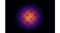
(Image credit: Harvard SEAS)
Recommended
Recommended
Read This Next
Read This Next
© Fraunhofer ILT, Aachen, Germany/Andreas Steindl
Photo 301002215 © Artitwpd | Dreamstime.com

(Image credit: Jerzy Szuniewicz)
Learning Resources
Learning Resources
Courtesy of Shanghai Optics Inc


Sponsored Content
(Image credit: Palashuddin Sk)

(Photocollage created by Jacob Deats, Laboratory for Laser Energetics)
(Image credit: Jiaye Chen)
Photo 21585186 © Image191 | Dreamstime.com

(Photo credit: Vescent)

(Photo credit: Quantum Science)

(Photo credit: QinetiQ)
(Image credit: VueReal)
Photo 305702414 © Zolak Zolak | Dreamstime.com

(Image credit: Focuslight Technologies/Susanne Westenhoefer)
(Image credit: Duke University)
(Image credit: Mikhail Volkov/University of Konstanz)
(Photo credit: ZEISS)
(Image credit: Endeavor Business Media)
(Photo credit: Quantum Corridor)

(Image credit: Duma Optronics)
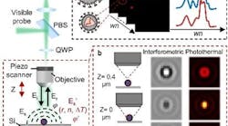
(Image credit: Ji-Xin Cheng’s Lab, Boston University)


(Image credit: TRUMPF Scientific Lasers)

(Image credit: EPFL/Gözden Torun)
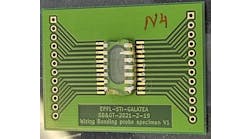
Photo 131219064 © Photoking | Dreamstime.com

Photo 174814076 © Artur Szczybylo | Dreamstime.com

Photo 11144854 © Mark Herreid | Dreamstime.com
(Image credit: GQI)
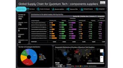
Photo 5322337 © Demarco | Dreamstime.com
(Image credit: Endeavor Business Media)
(Courtesy of JJ Coello-Bravo)

Featured Media
Featured Media
Home
Photonics Hot List: April 26, 2024
April 26, 2024

Home
Photonics Hot List: April 19, 2024
April 19, 2024
Home
Photonics Hot List: April 12, 2024
April 12, 2024

Leadership in Lasers and Photonics
Welcome to Laser Focus World's Leadership in Lasers and Photonics program.